
Lab 1 - Image 17
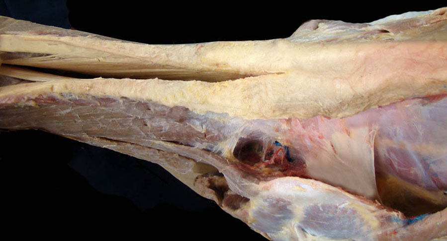
Dorsal view of equine withers region. The nuchal ligament is split on the midline and the left funicular part is labeled (1), cranially the lamellar part of the nuchal ligament is seen (2). Caudally the nuchal ligament merges with the supraspinous ligament which is not split (3). The dorsal part of the scapula (4) is displaced laterally to expose the dorsal scapular ligament (6). Also, part of the rhomboideus m. (5) has been removed to enhance exposure of the elastic dorsal scapular ligament (6).