Lab: Cerebellum
CLOSE
Canine Brain Transection
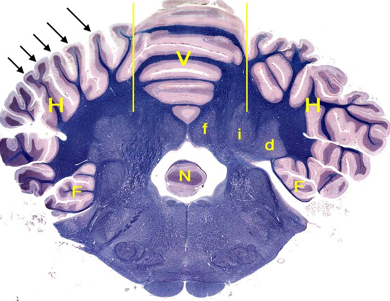
Transverse section through the cerebellum and medulla oblongata of a dog (luxol blue-crysl violet stain). The surface of the cerebellum is arranged into folia (black arrows). Each folium is bounded by sulci. Vertical yellow lines mark approximate boundaries between the central cerebellar vermis (V) and bilateral cerebellar hemispheres (H). The flocculus (F) is evident as a ventral lobule of the hemisphere. A profile of the nodulus (N), a caudoventral lobule of the vermis, is visible between confluent cerebellar peduncles connecting the cerebellum to the brainstem bilaterally. Three cerebellar nuclei can be seen deep within blue-stained white matter. From medial to lateral, the nuclei are: fastigial (f); interpositus (i); and dentate (d).
Go Top