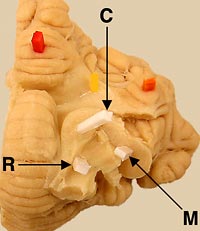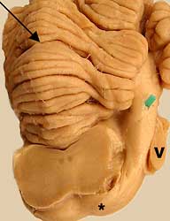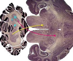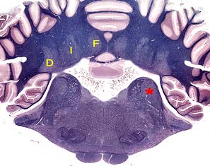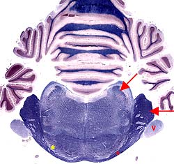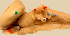
Bilaterally, three cerebellar peduncles connect the cerebellum to the brainstem. The peduncles are named for the directions they take in connecting to the brainstem (synonyms are shown below parenthetically).
- Caudal cerebellar peduncle (restiform body) connects the cerebellum to the medulla oblongata.
- Afferent fibers: The caudal peduncle contains cerebellar afferent fibers from the lateral cuneate nucleus and dorsal spinocerebellar tract, from vestibular nuclei and nerve, and from the olivary nucleus (climbing fibers).
- Efferent fibers: The caudal peduncle contains cerebellar efferent fibers from the fastigial nucleus (to reticular formation and vestibular nuclei), plus a small efferent contingent from flocculonodular cerebellar cortex (to vestibular nuclei).
- Middle cerebellar peduncle (brachium pontis) connects the ventral pons to the cerebellum.
- Afferent fibers: The middle peduncle is entirely afferent. The fibers come from neurons of pontine nuclei. Axons of the neurons form transverse pontine fibers that decussate and then collect dorsolaterally as the middle peduncle.
- Rostral cerebellar peduncle (brachium conjunctivum) connects the cerebellum to the midbrain and forebrain. Efferent fibers in the peduncle decussate immediately in the midbrain and thus they affect contralateral motor centers.
- Afferent fibers: The only afferents in the peduncle are from a ventral spinocerebellar tract (composed of projection axons that decussate within the spinal cord and then cross to the original side within the cerebellum).
- Efferent fibers: Axons from the interpositus nucleus synapse in the contralateral red nucleus, to influence activity in the rubrospinal tract. Axons from the dentate nucleus synapse in the contralateral thalamus, which projects to motor areas of cerebral cortex. (Note: Some efferent axons synapse on small neurons associated with the red nucleus; these small neurons project to the thalamus.)