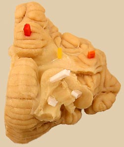
Surface Features:
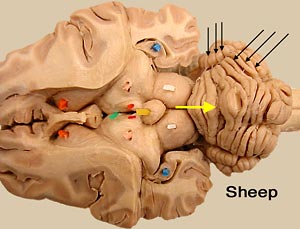
The surface of the cerebellum features folia (ridges) separated by sulci (grooves).
 Regions:
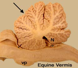
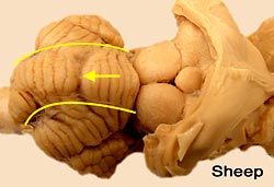
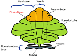
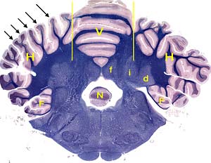
The central region of the cerebellum is designated vermis. It is flanked bilaterally by cerebellar hemispheres. Fissures divide the vermis into ten lobules. The primary fissure (the first fissure to develop embryonically) divides the cerebellum into rostral (anterior) and caudal (posterior) lobes.
The most caudal (caudoventral) lobule of the vermis is the nodulus. The most ventral lobule of each cerebellar hemisphere is called a flocculus. Bilateral flocculonodular tracts connect each flocculus to the nodulus. The combination constitutes a flocculonodular lobe, which is functionally related to vestibular nuclei.
 Gray Matter:
Cerebellar cortex refers to the gray matter covering the surface of the cerebellum. At low magnification, two of the three cortical layers are evident: a pale surface layer and a deep dark layer.
Three pair of cerebellar nuclei are present deep within cerebellar white matter. From medial to lateral, the nuclei are named: fastigial, interpositus, and dentate (lateral).
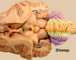 Zones:
Each cerebellar nucleus receives input from cerebellar cortex that is close to it. Thus, the cerebellum can be divided into three sagittal zones, based on cortical connections to nuclei.
From medial to lateral, the three longitudinal zones are commonly designated: vermis, paravermis, and hemispheres. (Alternatively, the three zones may be called: median, intermediate, and lateral.)