Brain Anatomy Introduction
CLOSE
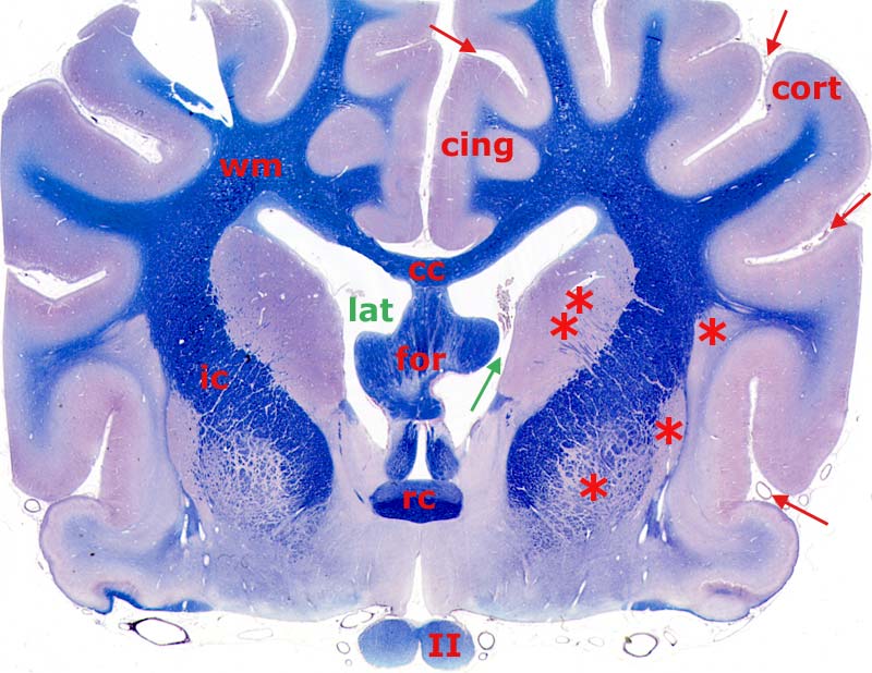
Canine Telencephalon Transection
Transverse section through the telencephalon of a canine brain (white matter is stained blue). Arrows indicate sulci (grooves), which delineate gyri. The cingulate gyrus (cing) is labeled. Gray matter on the surface of each cerebral hemisphere is cerebral cortex (cort). Deep gray matter constitutes basal nuclei (asterisks); the large medial basal nucleus is the caudate nucleus (double asterisk).
White matter (wm) of each cerebral hemisphere is continuous with internal capsule (ic) and corpus callosum (cc). The fornix (for), rostral commissure (rc) and optic nerve (II) are also labeled. The green arrow points to the interventricular foramen, which connects the third ventricle (below) with the lateral ventricle (lat). A piece of choroid plexus is evident dorsal to the right interventricular foramen.
Go Top