Brain Anatomy Introduction
CLOSE
Equine Caudal Brainstem
Dorsal view of an equine brainstem, minus the diencephalon. The three cerebellar peduncles are evident. The rostral peduncle (white) connects the cerebellum to the midbrain. The middle peduncle (red) connects the cerebellum to the ventral pons. The caudal peduncle (blue) connects the cerebellum to the hindbrain and spinal cord. The floor of the fourth ventricle (IV) is labeled.
In the myelencephalon, fibers of the caudal cerebellar peduncle (cp) pass deep to the dorsal cochlear nucleus (arrow) before ascending into the cerebellum (blue pic).
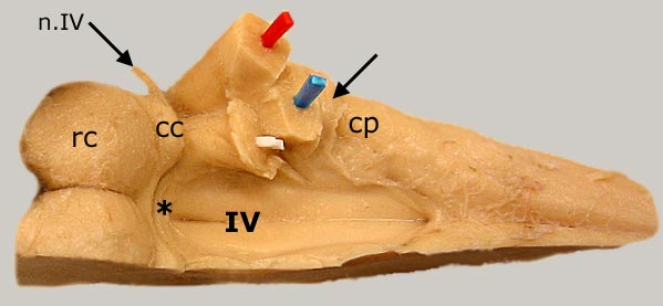
In the midbrain the caudal colliculus (cc) and the rostral colliculus (rc) are labeled. The trochlear nerve (n.IV) decussates (crosses sides) in the rostral medullary vellum (asterisk) and emerges from the dorsal surface of the brainstem.
Go Top