Brain Anatomy Introduction
CLOSE
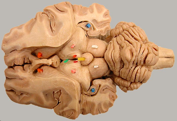
Sheep Diencephalon - Dorsal View
Dissection of sheep brain (dorsal cerebrum removed to expose the brainstem). The thalamus is marked bilaterally (pink pics). The green pick pokes through the third ventricle and into the interthalamic adhesion. The sheep pineal body (yellow) is large compared to that of the dog. Red picks mark habenular nuclei of the epithalamus. For a ventral view of the diencephalon, click here.
Some non-diencephalic structures are indicated: caudate nucleus (orange), hippocampus (blue), and rostral colliculus (white) of the midbrain.
Go Top