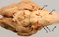
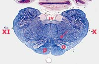
The myelencephalon (medulla oblongata) gives rise to seven cranial nerves (VI through XII) and contains most of the fourth ventricle. A characteristic feature of the myelencephalon is the presence of bilateral pyramids along the ventral surface. The pyramids contain fibers which arise in the motor cortex and travel to the spinal cord as corticospinal fibers (the fibers run through the internal capsule and crus cerebri and decussate at the brain-spinal cord junction).
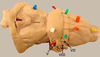
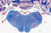
The rostral 25% of the medulla oblongata features: the trapezoid body and the dorsal nucleus of the trapezoid body. These structures are involved with hearing. The next 25% contains the facial nucleus and bilateral lateral foramina. The caudal 50% of the medulla oblongata can be recognized by the presence of a nucleus with a peculiar shape. The olivary nucleus is located just dorsal to the pyramid. The nucleus projects to the cerebellum and plays an important role in coordination of movement. Hypoglossal fibers from the hypoglossal nucleus exit by passing lateral to the olivary nucleus.
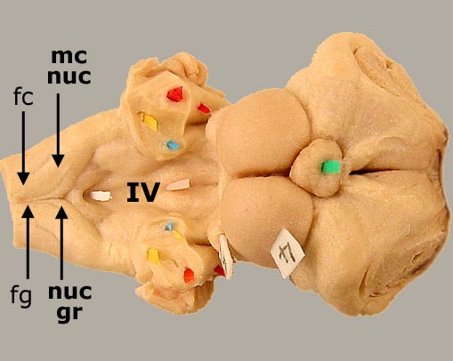
Two ascending tracts from the spinal cord terminate on nuclei in the caudal medulla oblongata: The fasciculus gracilis terminates in the nucleus gracilis . The fasciculus cuneatus terminates in the medial cuneate nucleus. These nuclei convey discriminative touch and kinesthesia information to the thalamus, which, in turn, projects to the somesthetic cerebral cortex. The fasciculus cuneatus also terminates and in a lateral cuneate nucleus which projects to the cerebellum.
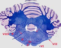
The thin roof of the fourth ventricle (caudal medullary vellum) gives rise to bilateral choroid plexuses. A choroid plexus is a highly vascular, villus structure that produces cerebrospinal fluid. A portion of each plexus passes through the lateral foramen of the fourth ventricle and spills into the subarachnoid space. An enlargement of the subarachnoid space, the cisterna magna, is a common site for obtaining cerebrospinal fluid.