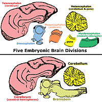
Recall that during embryonic development the brain is initially composed of three primary vesicles: Forebrain, Midbrain, and Hindbrain. These vesicles ultimately become five brain divisions: Telencephalon, Diencephalon, Mesencephalon (midbrain), Metencephalon, and Myelencephalon.
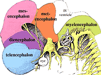
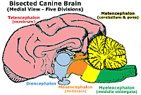
The five brain divisions are convenient for regionally categorizing the locations of brain components. After this lab, you should be able to identify brain divisions present in a brain section or gross brain specimen that you are asked to inspect.
| Embryonic Brain Division |
Derived Brain Structures |
Definitive Brain Cavities |
Associated Cranial Nerves |
| FOREBRAIN Telencephalon Diencephalon |
Cerebrum Thalamus, Hypothalamus,... |
Lateral ventricles Third ventricle |
Olfactory (I) Optic (II) |
| MIDBRAIN Mesencephalon |
Midbrain |
Mesencephalic aqueduct |
III & IV |
| HINDBRAIN Metencephalon Myelencephalon |
Pons & Cerebellum Medulla oblongata |
Fourth ventricle Fourth ventricle |
Trigeminal (V) VI - XII |
Note: The terms forebrain and hindbrain have clinical significance with respect to localizing lesions diagnostically in patients.
|
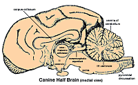
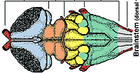
Another way to subdivide the brain is: cerebrum, cerebellum, and brainstem. |
|
|
The portion of brain remaining after the cerebrum and cerebellum are removed is referred to as the brainstem. Brainstem = diencephalon, mesencephalon, pons of the metencephalon, and myelencephalon. |
|
|
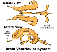
The embryonic neural cavity develops into a brain ventricular system. Each of the five brain divisions houses a ventricle, except for the mesencephalon which contains the mesencephalic aqueduct. The cerebrum contains two ventricles, one in each hemisphere. |
|