You may wish to perform the following dissection to supplement what you can observe in lab demonstration brains and textbook images. Do all dissection on the one-half sheep brain; preserve the whole sheep brain and half dog brain for surface features. Use a scalpel handle or penknife to make the cuts described below. Ask an instructor for assistance if the following instructions are not clear to you.
1. Separate the cerebral hemisphere from the brainstem:
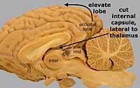
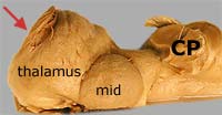
Lift the occipital lobe (caudal end) of the cerebral hemisphere to observe the thalamus of the brainstem. As you continue the lifting, cut the internal capsule in order to separate the cerebral hemisphere from the brainstem. (Refer to the following illustrations to locate the occipital lobe, internal capsule and thalamus.) Identify features of the exposed brainstem, including the rostral and middle cerebellar peduncles, which are more distinct than the caudal cerebellar peduncle.
2. Cerebral hemisphere dissection:
|
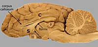
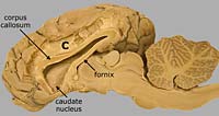
Scrape away cerebral cortex from the cingulate gyrus to observe corpus callosum joining the rest of the cerebral white matter. (The dissection procedure is illustrated in images to the right.) |
|
|
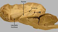
Remove the septum to expose the lateral ventricle. The caudate nucleus (the largest basal nucleus) lines the deep wall of the lateral ventricle at this level. Continue exploring the lateral ventricle by cutting between the corpus callosum and fornix. Look for a choroid plexus along the floor of the ventricle, just lateral to the fornix and fimbria. |
|
|
Remove the top of the cerebral hemisphere to expose the hippocampus, along the floor of the caudal half of the lateral ventricle. Axons from the hippocampus form the fimbria and fornix. Remove occipital lobe to trace the lateral ventricle and the hippocampus caudally and ventrally into the piriform lobe. |
|