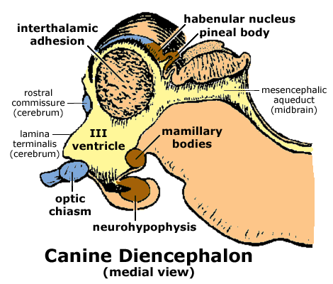
The diencephalon, which gives rise to the optic nerve, can be divided into four regions: thalamus, hypothalamus, subthalamus and epithalamus. A drawing of a median view of the rostral brainstem is shown below. Components of the diencephalon are labeled in bold.

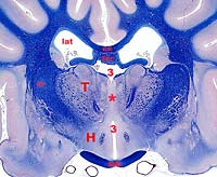
Most of the diencephalon is occupied by thalamus. The thalamus is composed of a large number of individual nuclei, each communicating with a region of cerebral cortex. All input to the neocortex relays first in the thalamus. The thalamus of right and left sides make contact at the midline, forming an interthalamic adhesion which obliterates the center space of the third ventricle.


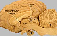
The third ventricle communicates with each lateral ventricle via an interventricular foramen. Third ventricle communicates with the fourth ventricle via the mesencephalic aqueduct.
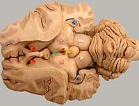
The lateral geniculate body (nucleus) is a thalamic nucleus that receives visual input via the optic nerve, optic chiasm (chiasma), and optic tract. Neurons of the lateral geniculate nucleus send axons via the internal capsule to the primary visual area of the cerebral cortex. The medial geniculate body (nucleus) receives auditory input and projects to the auditory cortex.
(Note: The term "geniculate body" is a surface term. The term "geniculate nucleus" refers to the same structure as seen in section. Medial and lateral geniculate bodies/nuclei are sometimes designated "metathalamus".)
The hypothalamus is visible on the ventral surface of the diencephalon, it includes the optic chiasm and mamillary bodies. Between these, the hypophysis (pituitary gland) is attached via a hollow stalk (infundibulum). The hypothalamus plays an important role in controlling endocrine and autonomic functions.