Spinal Cord Anatomy
CLOSE
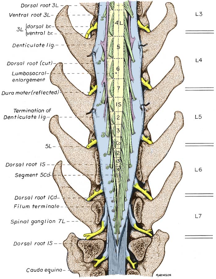
Canine Spinal Cord Termination
Illustration of a canine spinal cord termination. A laminectomy was performed and dura mater has been reflected to expose spinal cord segments and spinal roots. Dorsal roots are cut on the left side to expose the denticulate ligament (purple). Notice the termination of the denticulate ligament; the cauda equina; and the terminal filament (filum terminale), a glial continuation persisting beyond the functional end of the spinal cord. The illustrated position of the spinal cord termination at the L6-L7 intervertebral disc represents the most common relationship for medium and large dogs (varying plus or minus a half vertebra). In small dogs (under 7 kg) the typical position is one vertebra caudal to that shown.
Go Top