Spinal Cord Anatomy
CLOSE
Gray Matter Collateral Branches and Neurons
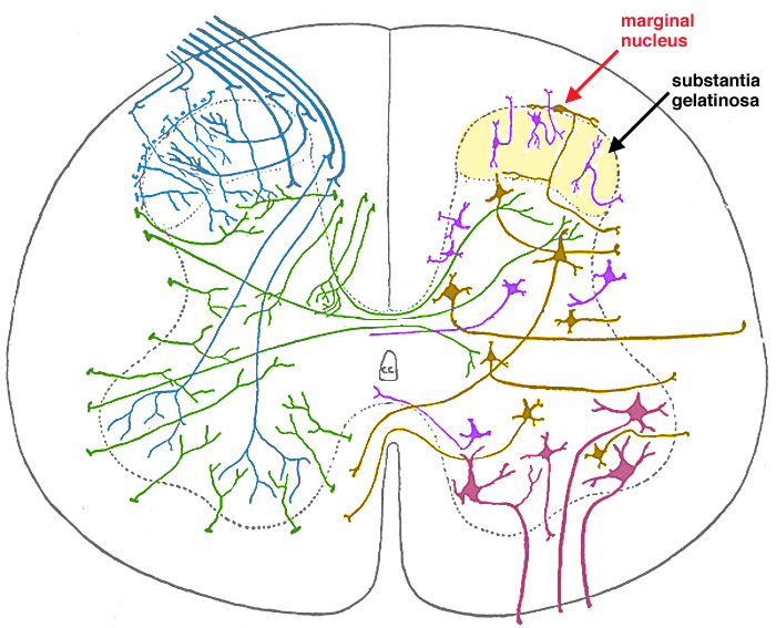
Drawing of a transverse section through a spinal cord segment. The left side shows collateral branches entering gray matter form white matter. The collateral branches are from cranial and caudal branches of primary afferent neurons (blue) or from spinal tracts (green) descending from the brain. The right side shows profiles of spinal neurons, which are either efferent neurons (maroon), projection neurons (brown), or interneurons (purple). The substantia gelatinosa (yellow) is a collection of small interneurons that appear to cap the dorsal horn of gray matter. The marginal nucleus, composed of flat neurons at the surface of the dorsal horn, is significant as a collection of pain projection neurons.

Go Top