Spinal Cord Anatomy
CLOSE
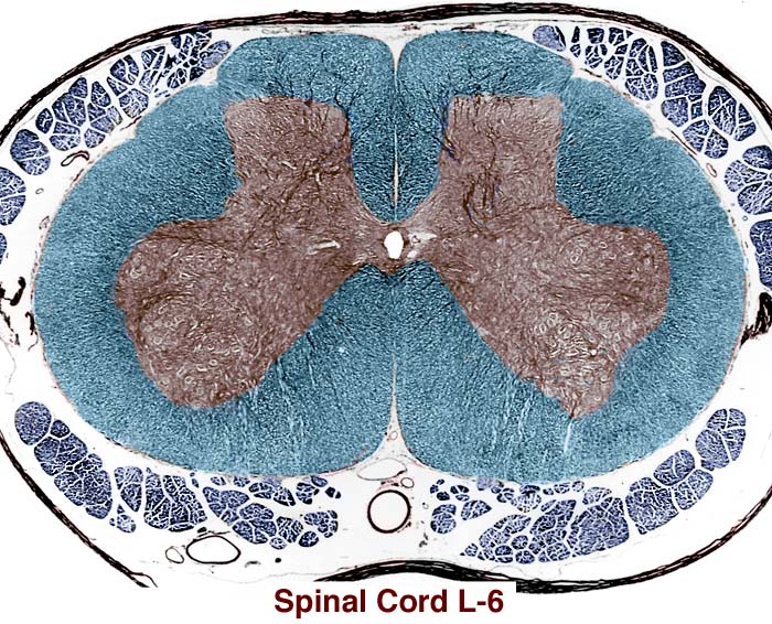
L6 Spinal Cord Segment
In this transverse section through the L6 segment of a canine spinal cord, please observe: The ventral median fissure is prominent, as are dorsolateral sulci. The dorsal median sulcus is continued by a dorsal median fissure, rather than a dorsal median septum. Identify the central canal and gray matter (brown). Gray matter dorsal horns are flat in the lumbosacral region. The lateral bulge in the ventral gray matter is produced by motor neurons that innervate the pelvic limb. White matter (blue) is less abundant than gray matter. The overall shape of the segment is oval. Spinal rootlets (dark blue profiles) are abundant. Pia mater, denticulate ligament, and dura mater can be seen; arachnoid is scarcely evident.
Go Top