Spinal Cord Anatomy
CLOSE
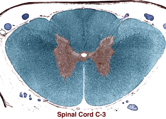
C3 Spinal Cord Segment
In this transverse section through the C3 segment of a canine spinal cord, please observe: The ventral median fissure is prominent, as are dorsolateral sulci. A dorsal rootlet is entering the right dorsolateral sulcus. Identify the central canal and gray matter (brown). Gray matter dorsal horns are pointed in the cervical region. The lateral bulge in the central gray matter is produced by neurons feeding spinal root of the accessory nerve. White matter (blue) is abundant. The overall shape of the segment is oval. Spinal rootlets (dark blue profiles) are sparse. Pia mater and dura mater can be seen; arachnoid is hardly evident.
Go Top