Spinal Cord Anatomy
CLOSE
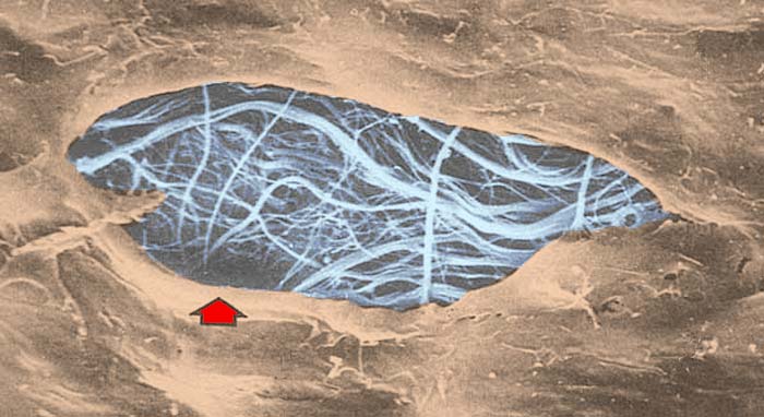
Pia Mater (SEM)
Below, is a scanning electron micrograph (SEM) of pia matter on the surface of the spinal cord. The view is from the subarachnoid space and there is a tear (red arrow) in the layer of flattened fibrocytes (brown) that line the entire subarachnoid space. Through the tear one can see the collagen fibrils (blue) that contact the glial-limiting-membrane (astrocyte processes) along the outer edge of the spinal cord. The flattened fibrocytes feature small processes, including cilia. The flattened fibrocytes are continuous with the perineural epithelioid cells associated nerves fascicles.
Go Top