All spinal pathways involve a sequence of neurons. Excitability is transmitted from one neuron to the next in the sequence. Pathways are either ascending (carrying information from receptors to the brain) or descending (conveying information from the brain to spinal cord neurons). Below, parts of a touch & kinesthesia ascending pathway and a corticospinal descending pathway are diagrammed.
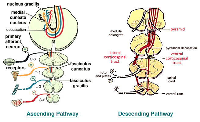
The initial neuron in an ascending spinal pathway is always a primary afferent neuron having a unipolar cell body in a spinal ganglion and receptors at the peripheral end of its axon (above left). The central end of its axon synapses on projection neurons within the spinal cord (or brain). In conscious pathways, projection neurons terminate in the contralateral thalamus, synapsing on a thalamic neuron that projects to the cerebral cortex.
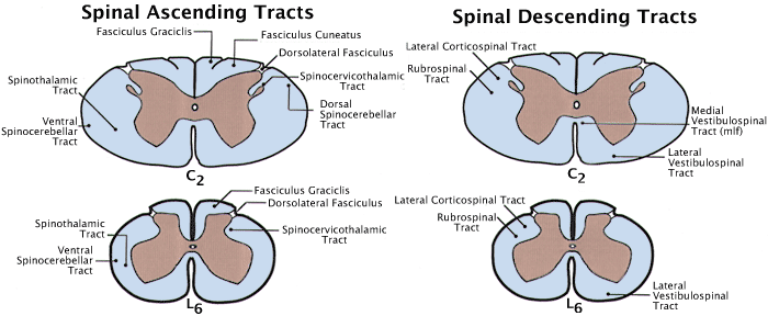
The above illustration shows approximate locations in spinal white matter of selective ascending tracts (left) and descending tracts (right). Tracts that decussate in the spinal cord are shown on the left side. Tracks that remain ipsilateral are shown on the right side. Cervical and lumbar levels of the spinal cord have different combinations of tracts because not all tracts run the whole length of the spinal cord. Descending tracts will be discussed latter in the course.
Selected Ascending Pathways
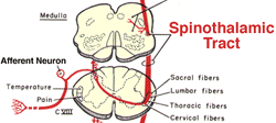 Pain and Temperature Ascending Pathway. These receptors are mainly free nerve endings. Axons of the primary afferent neurons are nonmyelinated (C-fibers) or thin myelinated axons (A-delta fibers) that bifurcate in the dorsolateral fasciculus. Projection neurons are located throughout the dorsal horn but they are concentrated in the marginal nucleus and nucleus proprius. Axons of the projection neurons decussate and ascend in the spinothalamic tract to reach the contralateral thalamus. Thalamic projection neurons send axons through the internal capsule to the somesthetic area of the cerebral cortex.
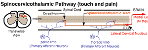 In carnivores, the spinocervicothalamic pathway is an important pain pathway. The pathway is unusual because it has two spinal projection neurons (and four instead of the usual three neurons for conscious pathways). The first projection neuron runs from the dorsal horn to the ipsilateral lateral cervical nucleus (located only in segments C1 & 2). The axon of the second projection neuron, arising in the lateral cervical nucleus, decussates and runs to the contralateral thalamus. Thalamic neurons project to the somesthetic area of the cerebral cortex.
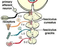 Touch and Kinesthesia Ascending Pathway. The pathway begins with encapsulated mechanoreceptors associated with primary afferent neurons featuring myelinated axons and large unipolar cell bodies. The axons bifurcate in the dorsal funiculus and ascend in fasciculus gracilis (pelvic limb and trunk) or fasciculus cuneatus (thoracic limb and neck) to, synapse in, respectively, nucleus gracilis or medial cuneate nucleus in the brainstem. From these nuclei, axons of projection neurons decussate (cross) and run in the medial lemniscus to the contralateral thalamus. Thalamic projection neurons send axons through the internal capsule to the somesthetic area of the cerebral cortex.
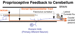 Subconscious Spinocerebellar Pathways. Subconscious pathways typically are ipsilateral (uncrossed) and involve only two neurons (primary afferent & projection neurons). In the case of spinocerebellar pathways, which you will study later in this course, receptors are mainly encapsulated proprioceptors associated with large myelinated axons, since the information is generally urgent. Pelvic and thoracic limb spinocerebellar pathways differ. Primary afferent neurons from the pelvic limb synapse on the nucleus thoracicus, which gives rise to the dorsal spinocerebellar tract. For the thoracic limb, cranial branches of primary afferent neurons run in the fasciculus cuneatus and synapse in the lateral cuneate nucleus. Axons of projection neurons from both limbs run in the caudal cerebellar peduncle to reach the cerebellum.