Spinal Cord
In transverse section, the spinal cord features:
�
-- a central canal lined by ependymal cells;
�
-- gray matter (butterfly-shaped profile) surrounding the central canal; and
�
-- white matter surrounding the gray matter; also notice,
�
-- spinal roots and meninges external to the spinal cord.
Examine glass slide 54 in your Histology slide box, lumbar spinal cord transection from a dog, or Neuroanatomy glass slide A-1, canine spinal cord transections from four segments (H&E stain). Identify the spinal cord features illustrated below:
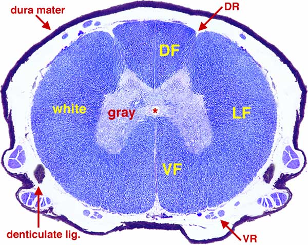 �
�Fig. 13.Canine spinal cord segment T-6. At low power, identify H-shaped gray matter surrounding the central canal (asterisk). The dorsal and ventral projections of gray matter are called "horns". Notice that ventral horns are broader than dorsal horns (particularly in segments that innervate limbs) and that this thoracic segment has small lateral horns. White matter (DF/LF/VF) is exterior to gray matter and spinal roots (DR/VR) and meninges (dura mater & denticulate lig.) are external to the spinal cord.
At higher power, further examine the spinal cord transection:
Find ependymal cells lining the central canal. Examine white matter (where neuron perikarya are absent) for two types of neuroglial cells, astrocytes and oligodendrocytes, distinguished by their nuclei (see Fig. 6). Examine ventral horn gray matter at high power to find large multipolar neurons (see Fig. 14) and observe meninges (Fig. 15).
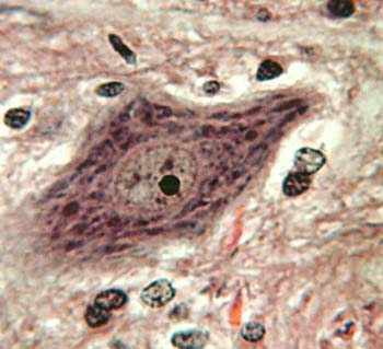 |
Fig. 14. H&E stained spinal gray matter exhibits a variety of perikarya belonging to neurons and glial cells. Examine ventral horn gray matter at high power to find perikarya of the large motor neurons that innervate skeletal muscle. These multipolar neurons have a leptochromatic nucleus, a prominent nucleolus, and distinct clumps of Nissl substance (RER) in the cytoplasm of the cell body and proximal dendrites. |
 �
��
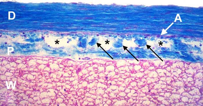
Fig. 15. Meninges are connective tissue layers surrounding the brain, the spinal cord, and the roots of peripheral nerves. The pia mater (P) is a delicate connective tissue layer attached directly to the white matter of the spinal cord. The dura mater (D) is a thick connective tissue layer. It is the most superficial of the three meningeal layers. The arachnoid (arachnoid membrane) is attached to the deep surface of the dura mater (A). Arachnoid trabeculae (arrows) extend from the arachnoid to the pia mater. The subarachnoid space (asterisks), between the arachnoid and pia mater, is lined by flat fibrocytes and contains cerebrospinal fluid. (The subarachnoid space is collapsed due to fluid loss in the above image.)
�
Go Top