Neurons
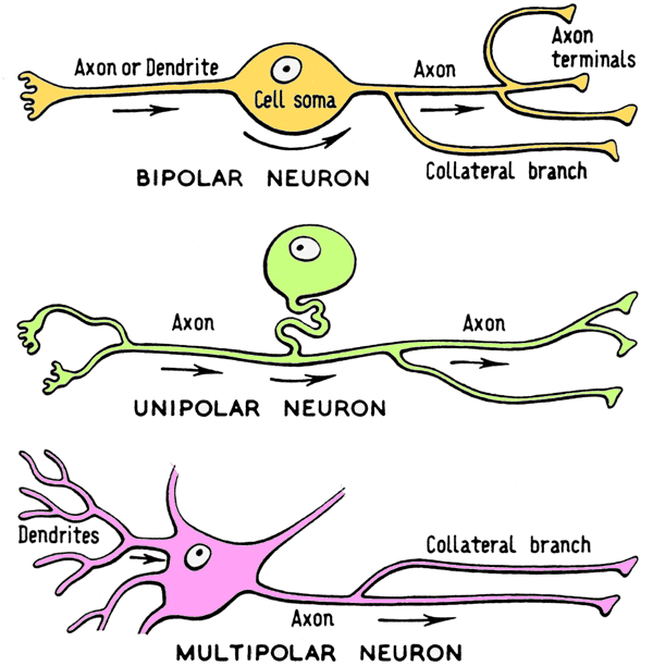
Three major categories of neurons are recognized:
Bipolar neurons are relatively rare. They are sensory neurons found in olfactory epithelium, the retina of the eye, and ganglia of the vestibulocochlear nerve.
Unipolar (pseudo-unipolar) neurons are sensory neurons with cell bodies located in spinal and cranial nerve ganglia. (Note: unipolar neurons are sometimes called pseudo-unipolar because embryologically they originate as bipolar neurons and subsequently become unipolar.)
Multipolar neurons are the most common type of neuron. They are located in the central nervous system (brain and spinal cord) and in autonomic ganglia. Multipolar neurons have more than two processes emanating from the neuron cell body.
 �
�Multipolar neurons:
Search for multipolar neurons in glass slide 48 in your Histology slide box, cerebellar cortex of pig (Golgi stain).
Note: Three layers are recognized in the cerebellar cortex (superficial gray matter of the
cerebellum):
(1) an outer molecular layer composed of few cells and many nonmyelinated fibers,
(2) an intermediate layer comprised of flask-like cell bodies called Purkinje cells,
which are multipolar neurons, and
(3) an inner granular layer composed of tightly-packed cells and fibers.
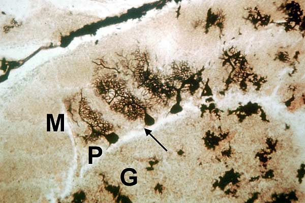
On glass slide 48, find a section similar to that shown below.
 �
�At middle of the slide, cell bodies of three Purkinje cells (neurons) are visible as black, rounded, profiles. Cell processes, termed dendrites, extend superficially from the cell body into the molecular layer. A small axon (arrow) emerges from each cell body and runs through the granue cell layer. (Note: Axons and dendrites of neurons are normally visible only in special stained sections such as this Golgi preparation.)
Question: Why is the Purkinje cell considered a multipolar neuron?
All multipolar neurons have two characteristics:
-- more than two processes emanate from the cell body, and
-- the cell body receives synaptic input just like the dendrites
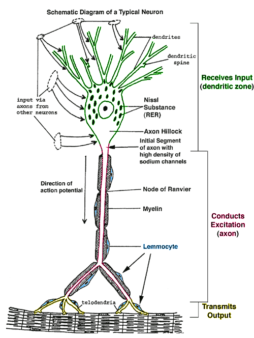
Somatic Efferent Neuron:
 �
��