CNS - Image 10
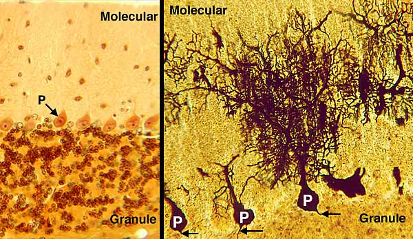
The cerebellar cortex is shown with two stains. Left A special silver stain shows cell bodies of all cortical neurons. Right A Golgi stain selectively stains Purkinje neurons. The molecular layer of the cerebellar cortex has few cell bodies but lots of dendrites of Purkinje neurons. The Purkinje cell layer is formed by cell bodies (P) of Purkinje neurons. Purkinje axons (arrows) run deep into white matter. The granule cell layer is densely populated with very small neurons.