|
|
|
 |
|
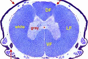
CNS - Image 1:
Transverse section through the T-6 spinal cord segment (Luxol Blue stain). The central canal (asterisk) is surrounded by butterfly-shaped gray matter. Blue stained white matter is superficial to the gray matter. |
|
|
|
 |
|
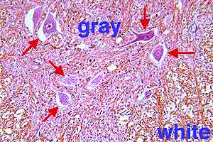
CNS - Image 2:
Spinal gray matter and white matter (H&H stain). Gray matter contains neuron cell bodies (arrows) along with glial cells and some myelinated fibers. White matter features dense accumulations of myelinated fibers and an absence of neuron cell bodi. |
|
|
|
 |
|
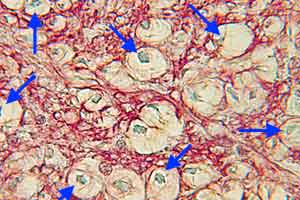
CNS - Image 3:
High power view of spinal white matter (special stain). Axons (arrows) of myelinated fibers can be seen within vacuoles produced by extraction of myelin lipid during tissue processing. Myelin neurokeratin (red) and glial nuclei are visible |
|
|
|
 |
|
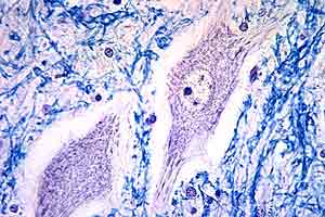
CNS - Image 4:
High power view of spinal gray matter (Luxol Blue &s Hematoxylin stain). Profiles of two cell bodies of large multipolar neurons are featured. Smaller neurons, glial cells, and neurokeratin of myelinated fibers are scattered throughout the background. |
|
|
|
 |
|
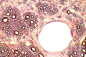
CNS - Image 5:
Intermixed regions of gray and white matter plus dilated vessels (myelin stain on a thin plastic section). Islands of myelinated fiber profiles (dark rings) are set in a gray matter background.
|
|
|
|
 |
|
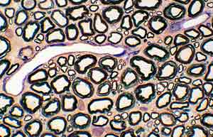
CNS - Image 6:
Spinal white matter (Wolter myelin stain on a thin plastic section). Myelin sheath profiles appear as dark rings. A vessel runs longitudinally through the section.
|
|
|
|
 |
|
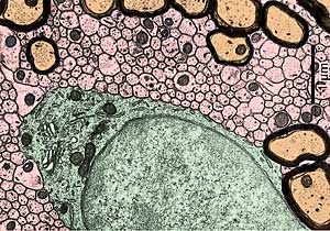
CNS - Image 7:
Electron micrograph of white matter in rat in spinal cord. Profiles of non-myelinated axons are colored pink. Notice that the non-myelinated axons are bare.
|
|
|
|
 |
|
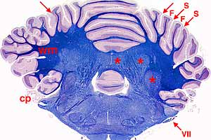
CNS - Image 8:
Transverse section through the hindbrain (Luxol Blue & Hematoxylin stain). Situated above the brainstem, the cerebellum features deep gray matter called nuclei (asterisks), white matter (wm), and superficial gray matter called cerebellar cortex. |
|
|
|
 |
|
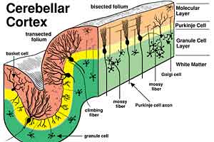
CNS - Image 9:
An artist drawing of the cytology and circuitry of the cerebellar cortex is presented. The outer molecular layer (brown) has dendrites and axons but few cell bodies. The Purikinje cell layer (yellow) contains large Purkinje (piriform) neurons. The granule cell layer (green) contains a dense accumulation of very small neurons. |
|
|
|
 |
|
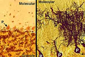
CNS - Image 10:
The cerebellar cortex is shown with two stains. The molecular layer of the cerebellar cortex has few cell bodies but lots of dendrites of Purkinje neurons. The Purkinje cell layer is formed by cell bodies (P) of Purkinje neurons. The granule cell layer is densely populated with very small neurons.
|
|
|
|
 |
|
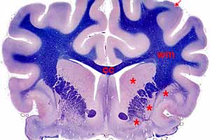
CNS - Image 11:
Transverse section through the cerebrum of the forebrain (Luxol Blue & Hematoxylin stain). The cerebrum features deep gray matter called nuclei (asterisks), white matter (wm), and superficial gray matter called cerebral cortex.
|
|
|
|
 |
|
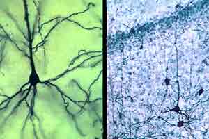
CNS - Image 12:
Cerebral pyramidal neurons are shown at two magnifications (Golgi stain). Cell bodies of pyramidal neurons have a pyramid shape with a single apical dendrite and multiple basal dendrites.
|