
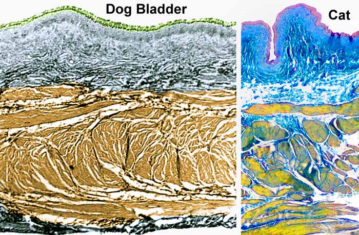
Above: Tissue sections through the bladder wall of a dog and cat are shown with color added. The detrusor muscle (smooth muscle coat of the bladder apex and body) has muscle fascicles variably organized rather than arranged in consistent layers. When empty, bladder mucosa exhibits folds that disappear during distension. As the bladder distends, the muscle coat becomes thinner and individual muscle fascicles shift position relative to one another. (With major distension the direction of detrusor fascicles shifts from an encircling orientation toward a tangential one, which presents a mechanical disadvantage.)
Bladder mucosa consists of a transitional epithelium (urothelium) resting on a lamina propria (top). A capillary plexus is present immediately deep to the epithelium (not evident here). A submucosa is between the mucosa and muscle coat. (Presence of a muscularis mucosae between lamina propria & submucosa is inconsistent and not evident here).
Below: The urinary bladder (like ureter and cranial urethra) is lined by transitional epithelium. Non-distended surface epithelial cells appear plump (left; arrows) compared to epithelial cells somewhat stretched (right).
The urothelium is bacteriostatic by virtue of a glycosaminoglycan secretion that impairs bacterial adhesion to the epithelium. During cystitis, molecules released by urothelium affect afferent nerve endings, producing bladder pain and hyperactivity.