
| Home Page | Embryology | Anatomy | Innervation | Physiology | Clinic Case |
 |
 |
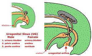
The embryonic hindgut terminates in a cloaca from which the allantois arises and into which mesonephric ducts empty. A urorectal septum divides the cloaca into, dorsally, a rectum-anal canal and, ventrally, a
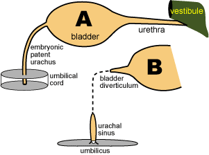
urogenital sinus which gives rise to the urinary bladder and urethra. The embryonic bladder is connected to the allantoic cavity by the urachus, which can be a postnatal problem if it persists as a patent duct or as a umbilical sinus or as a vesical diverticulum.
 The cranial urogenital sinus becomes urinary bladder. The mid urogenital sinus becomes pelvic urethra. The caudal urogenital sinus becomes the vestibule and vagina in females and penile urethra in males.
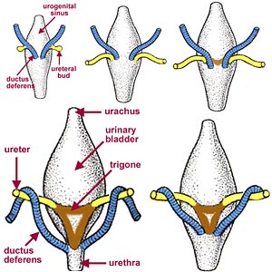
Bilaterally each mesonephric duct sprouts a ureteric bud just before terminating in the mid urogenital sinus. Each ureteric bud gives rise to a ureter and definitive kidney. As the urogenital sinus enlarges, it engulfs the ureters so they and the mesonephric ducts (future ducti deferentes) open separately into the sinus.
Ureters undergo repositioning in the urogenital sinus. They shift cranially so they ultimately open anterior to the bladder neck sphincter. The repositioning produces the trigone and urethral crest which serve to anchor the both ureters. Failure to properly reposition can result in an ectopic ureter, a source of postnatal incontinence when it opens distal to sphincters in the caudal urethra or vagina.