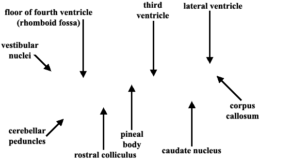
Dissected Equine Brain — Dorsal View


Ventricles are exposed in this dorsal view of a dissected equine brain. The lateral ventricle is the space medial to the caudate nucleus (1). The medial wall of the ventricle is thin between the corpus callosum and fornix. The third ventricle is evident anterior to the pineal body (green pic). The floor of the flourth ventricle (rhomboid fossa) is marked by white pics. Click here for another view.
LABELS
Go Top
RETURN