Equine Brain — Median View
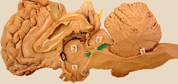
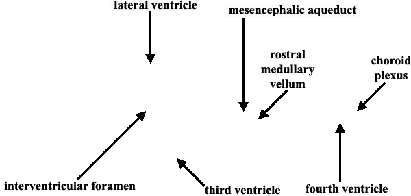
Components of the ventricular system are visible in this median view of a dissected equine brain. The medial wall of the lateral ventricle (septum pellucidum) has been torn between the corpus callosum and fornix, exposing the lateral ventricle. The third ventricle (4) surrounds the interthalamic adhession (3). Lamina terminalis (2) forms the rostral wall of the third ventricle. A green pic marks the mesencephalic aqueduct (ventral to rostral colliculus (6)). The fourth ventricle (5) is evident ventral to the cerebellum. The roof of the foruth ventricle is formed by rostral medullary vellum and caudal medullary vellum, the latter gives rise to choroid plexus. Click here for another view.
LABELS
Go Top
RETURN