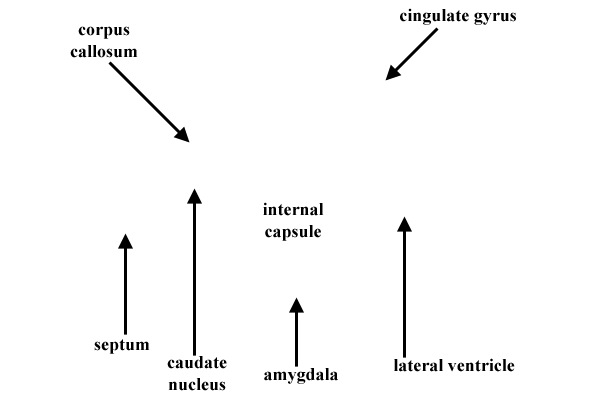
Lateral Ventricle in Sheep Cerebral Hemisphere


The hippocampus and fornix have been removed in this isolated cerebral hemisphere, to show the lateral wall of the lateral ventricle. The caudate nucleus can be seen rostrally in the wall of the ventricle. Ventral to the nucleus, the septal region forms a medial wall of the ventricle. The corpus callosum is evident dorsal to both the caudate nucleus and lateral ventricle. The lateral ventricle extends caudoventrally into the piriform lobe. For the other image, click here.
LABELS
Go Top
RETURN