Dissected Sheep Brain — Dorsal View
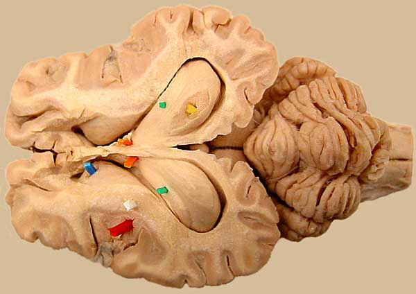
The top half of the telencephalon has been removed to reveal lateral ventricles bilaterally. The hippocampus (yellow pic), fimbria (green) and fornix (orange) form a floor for the lateral ventricle at this level. In the left hemisphere, cerebral cortex (red), internal capsule (white), and caudate nucleus (blue) are marked. Corpus callosum unites the two hemisphers along the midline. For the other image, click here.
LABELS
Go Top
RETURN