Equine Brain — Median View
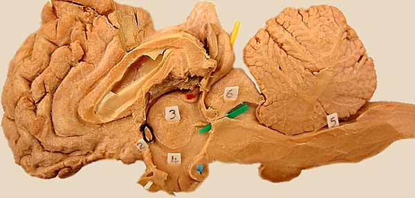
The right half of a dissected equine brain is showm in median view. The dorsocaudal part of the telencephalon and the medial wall of the lateral ventricle have been ablated. The lateral ventricle is visible between the corpus callosum and fornix. (Labeled diencephalics structures include: habenular nucleus (red pic), interthalamic adhesion (3), third ventricle (4), optic chiasm (white), and mamillary body (blue). For another image, click here.
LABELS
Go Top
RETURN