Dissected Sheep Brain — Dorsal View
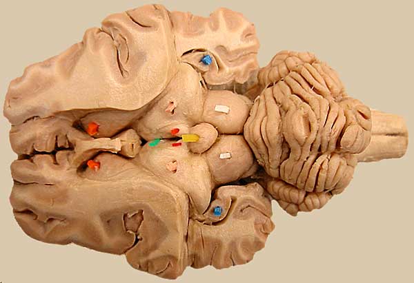
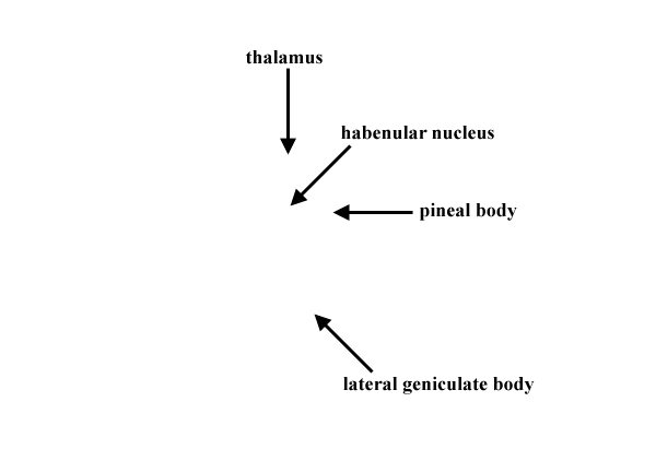
The telencephalon has been dissected to reveal the underlying diencephalon, including: thalamus (pink), habenular nuclei (red), pineal body (yellow), and third ventricle (green). Also labeled bilaterally are: midbrain rostral colliculus (white) and hippocampus (blue) and caudate nucleus (orange) of the telencephalon.
LABELS
Go Top
RETURN