Sheep Brain — Median View
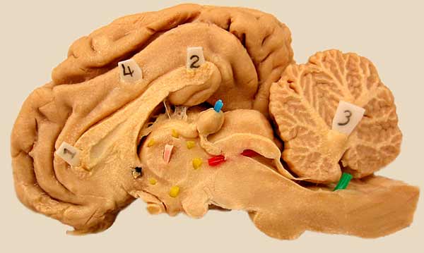
The right half of a dissected sheep brain is shown in median view. In the diencephalon, the interthalamic adhesion (pink), pineal body (blue), and thrid ventricle (yellow) are marked. The red pic marks the mesencephalic aqueduct. A green pic marks the fourth ventricle.
LABELS
Go Top
RETURN