Anatomy of the Dog Images
Brain Chapter 18
http://vanat.cvm.umn.edu/brain18/
This Page Displays A Sampling of New Images
by T. F. Fletcher
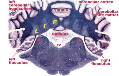
Cerebellar Nuclei
|
Click the image to view an enlarged version and caption in a separate window.
Fig. 46
|
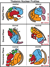
Thalamic Nuclei
|
Click the image to view an enlarged version and caption in a separate window.
Fig. 23
|
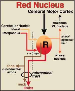
Red Nucleus
|
Click the image to view an enlarged version and caption in a separate window.
Fig. 20
|
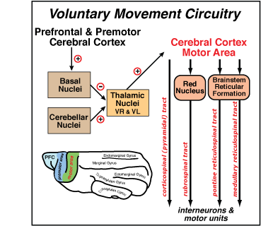
Vountary Movement Circuit
|
Click the image to view an enlarged version and caption in a separate window.
Fig. 43
|
Sulci & Gyri
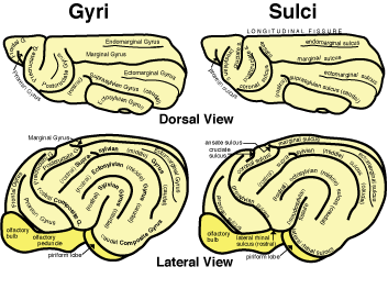
|
Click the image to view an enlarged version and caption in a separate window.
Fig. 30
|
Cranial Nerve Nuclei
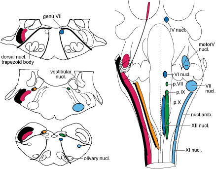
|
Click the image to view an enlarged version and caption in a separate window.
Fig. 5
|
Cerebellar Connections

|
Click the image to view an enlarged version and caption in a separate window.
Fig. 49
|
Limbic System Components
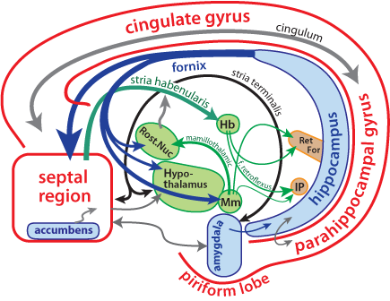
|
Click the image to view an enlarged version and caption in a separate window.
Fig. 39
|
Brainstem Reticular Nuclei
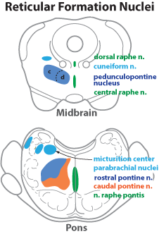
|
Click the image to view an enlarged version and caption in a separate window.
Fig. 6
|
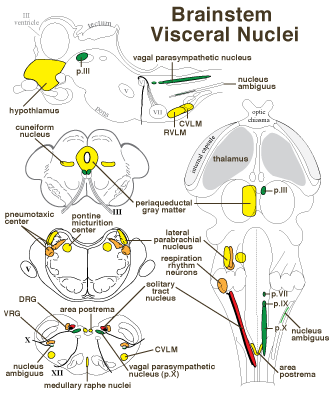
Brainstem Visceral Nuclei
|
Click the image to view an enlarged version and caption in a separate window.
Fig. 7
|
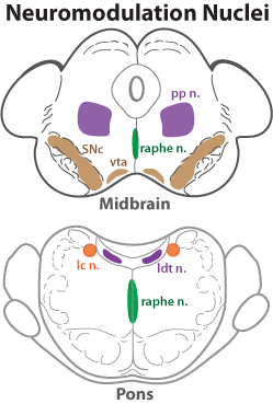
Brainstem Neuromodulatory Nuclei
|
Click the image to view an enlarged version and caption in a separate window.
Fig. 15
|
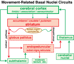
Basal Nuclei

|
Click the image to view an enlarged version and caption in a separate window.
Fig. 44
|
Basal Nuclei
|
Click the image to view an enlarged version and caption in a separate window.
Fig. 45
|
Hypothalamic Nuclei

|
Click the image to view an enlarged version and caption in a separate window.
Fig. 25
|
Cerebellar Lobes

|
- Click the image to view an enlarged version and caption in a separate window.
Fig. 47
|
Embryonic Brain Divisions

|
- Click the image to view an enlarged version and caption in a separate window.
Fig. 1
|
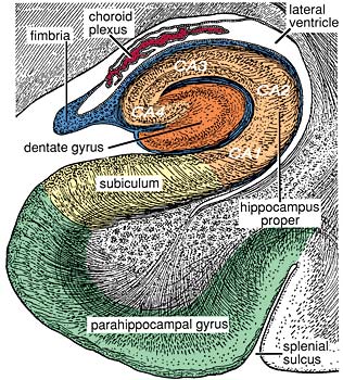
Hippocampal Formation
|
- Click the image to view an enlarged version and caption in a separate window.
Fig. 37
|
Neocortical Layers
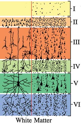
|
- Click the image to view an enlarged version and caption in a separate window.
Fig. 41
|
Cortical Regions

|
- Click the image to view an enlarged version and caption in a separate window.
Fig. 42
|
Cerebellar Folium.
 |
- Click the image to view an enlarged version and caption in a separate window.
Fig. 50
|
Brain Transection: Optic Chiasm

|
Click the image to view an enlarged version and caption in a separate window.
Fig. 24
|
Go Top