
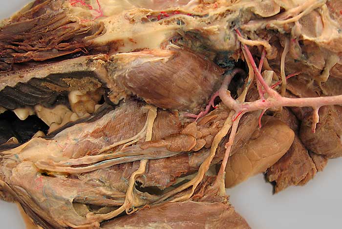
The tongue has been reflected ventrally and mucosa has been removed in this medial view of the right side of the head. The lingual n. (1), from the mandibular division of the trigeminal n., can be seen running on the mylohyoid m. (2). The nerve gives off a sublingual branch (3) before terminating as the sensory nerve of the tongue. The hypoglossal n. (4) can be seen passing lateral to the external carotid a. to innervate muscles of the tongue. The lingual artery (5), a branch of the external carotid a., accompanies the nerve.
Notice also: styloglossus m. (6); medial pterygoid m.(7); digastricus m. (8); mandibular salivary gland (9); polystomatic sublingual salivary gland (10); and mandibular & sublingual salivary ducts (asterisks).