Lab 24 - Image 6


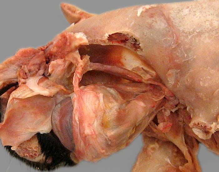
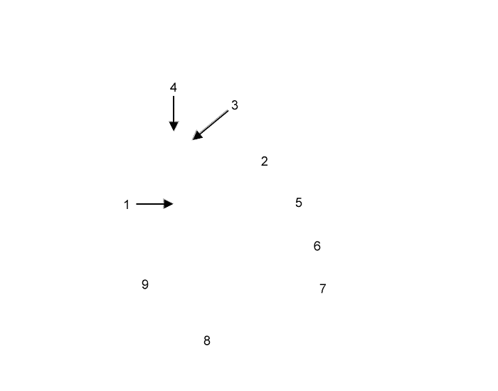
Dorsal view of the orbit of a cat with periorbita removed and the eyebll pulled laterally. The tendon (1) of the dorsal oblique muscle (2) hooks around the cartilaginous trochlear (3), which is anchored to bone by ligaments (4). Notice the medial rectus m. (5), dorsal rectus m. (6), and lateral rectus m. (7). The sclera (8) and cornea (9) of the eyeball are evident.
