
Lab 24 - Image 1
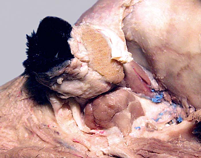
Lateral view of the orbit following removal of the periorbita to expose extrinsic eye muscles (1). The lacrimal gland (2) is exposed, deep to the orbital ligament (3) that runs from the zygomatic process (4) of the frontal bone to the previously removed zygomatic arch. The zygomatic salivary gland (5) is ventral to the orbit. The temporal m. has been removed from the temporal fossa (6), exposing the orbit and vessels (7) on the surface of the pterygoid m. (8).