
Lab 23 - Image 7
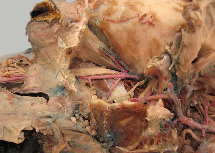
Deep dissection. The temporal m. has been removed, exposing the osseous temporal fossa (1). The top of the ramus of the mandible (2) has been cut off and the ramus has been reflected laterally, disarticulating the temporomandibular joint (asterisks). Remains of the masseter m. are evident on the lateral surface of the ramus.
The maxillary a. & n. (3) run on the surface of the medial pterygoid m. (4). The maxillary a. emerges from an alar canal (5) and runs through an infraorbital canal to emerge as an an infraorbital a. (6). The zygomatic salivary gland (7) is located ventral to the orbit which is enclosed within periorbita (8).