
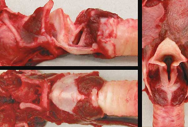
Three views of the larynx (fresh tissue). Right: epiglottic cartilage (1), arytenoid cartilages (2), aryepiglottic fold (3), thyroid cartilage (4), cricoid cartilage (5), cricoarytenoideus dorsalis m. (6), trachea (7), and root of the tongue (8).
Left top: The left side of the larynx has been removed. Identify: thyroid cartilage (1), epiglottic cartilage (2), arytenoid cartilage (3), cricoid cartilage (4), cricoarytenoideus lateralis m. (5), cricoarytenoideus dorsalis m. (6), basihyoid bone (7), and thyrohyoid bone (8). Notice the vocal ligament (9) and vocalis m. (10) of the vocal fold (covering mucosa removed). The laryngeal ventricle (11) is just rostral to the vocal fold.
Left bottom: thyroid cartilage (1), cricoid cartilage (2), cricothyroid ligament (asterisk), cricothyroideus m. (3), trachea (4), basihyoid bone (5), and ceratohyoid bone (6).