
Lab 23 - Image 13
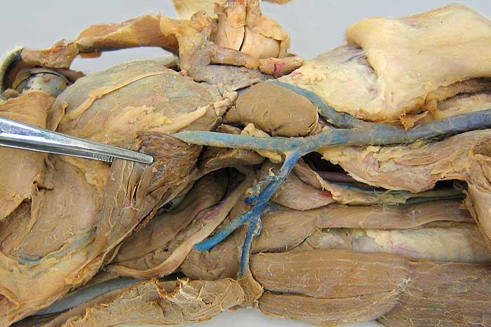
Ventral view of the left half of the head and neck. The digastricus m. (1) is reflected to expose the styloglossus m. (2), hypoglossal n. (3) and the hyoglossus m. (4). The latter attaches to hyoid bones, as does: sternohyoideus m. (5), geniohyoideus m. (6) and thyrohyoideus m. (7). Identify the trachea (8), sternothyroideus m. (9), medial retropharyngeal lymph node (10), mandibular salivary gland (11), monostomatic sublingual salivary gland (12), and the parotid salivary gland (13).