
Lab 21 - Image 6
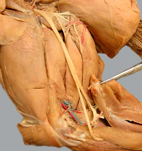
Lateral view of a cat pelvic limb with the biceps femoris m. (1) reflected. The sciatic n. (2) can be seen sending branches (3) to innervate hamstring muscles and then dividing into a common fibular (peroneal) n. (4) to cranial muscles of the crus and a tibial n. (5) to caudal muscles of the crus. The soleus m. and the lateral head of the gastrocnemius m. are reflected (6) to expose tibial n. branches (7) to caudal crus muscles.
Also notice: quadriceps femoris m. (8), adductor m. (9), semimembranosus m. (10) and semitendinosus m. (11). The popliteal a. (12) is visible.