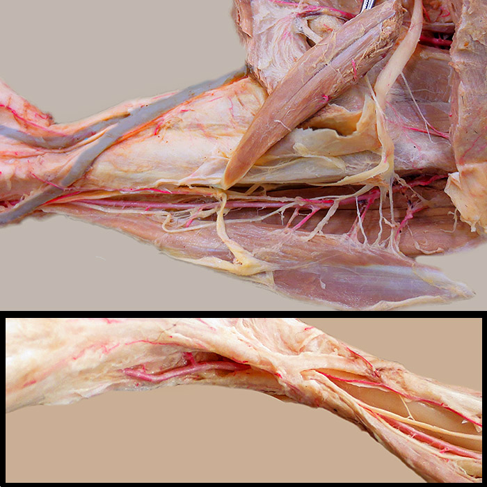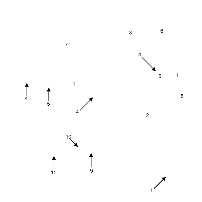

Above: The cranial tibial a. (1) has been exposed by reflecting the cranial tibial m. (2) and the fibularis longus m. (3) in the crus of a dog. Superficial (4) and deep (5) branches of the common fibular nerve (6) are exposed. (The superficial branch was accidentally cut.) Also evident are: cranial and caudal branches of the medial saphenous v. (7) and the long digital extensor m. (8).
Below: The insert shows that the cranial tibial a. (1) is renamed dorsal pedal a. (9) in the pes. The dorsal pedal a. ends in an arcuate a. (10) that gives rise to dorsal metatarsal aa. The largest of these is dorsal metatarsal a. II, the most medial. The main flow is directed to the plantar surface via the perforating branch (11) of dorsal metatarsal a. II.
