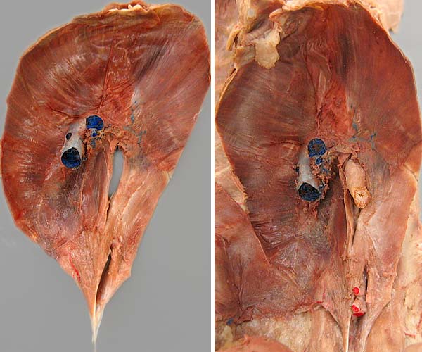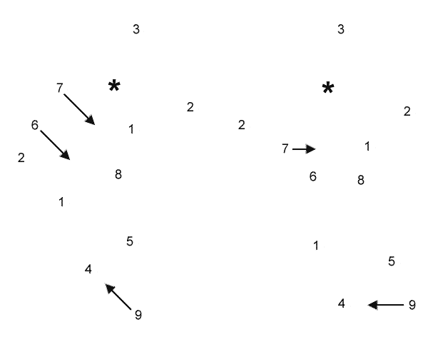

The diaphragm (viewed from the abdomen with ventral at the top) is shown removed from the cadaver (left) and in situ (right). The diaphragm has a horse-shoe shaped tendinous center (1) that separates an outer rim of skeletal muscle from muscular crura. The outer diaphragmatic muscle can be divided into costal (2) and sternal (3) regions. Dorsal to the tendinous center, notice that the right crus (4) and the left crus (5) have tendons (which attach to the bodies of lumbar vertebrae).
The tendinous center region contains a caval foramen through which the caudal vena cava (6) passes (also a hepatic vein (7) joining the caudal vena cava and the falciform ligament (asterisk) are evident). Between the crura, the esophagus passes through the esophageal hiatus (8) and the aorta passes through the aortic hiatus(9).
