
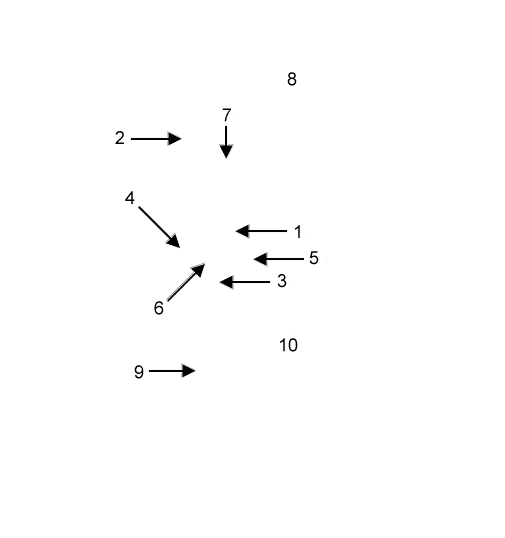
Lateral view of the scapular region and brachium. The axillary a. (1) gives off a large subscapular a. (2) which runs dorsally along the caudal border of the scapula. After giving rise to a cranial circumflex humeral a. (not visible), the axillary a. becomes the brachial a. (3). The subscapular a. gives rise to the thoracodorsal a. (4) and the caudal circumflex humeral a. (5). The latter forms an arterial loop around the humerus in conjunction with the cranial circumflex humeral a. (6) which is a terminal branch of the axillary a.
Two nerves pass from medial to lateral in the brachium: The axillary n. (7) can be seen innervating the reflected deltoideus m. (8). The radial n. (9) runs normally on the caudal surface of the brachialis m. (10). The nerve is pulled away from the muscle in this dissection.