
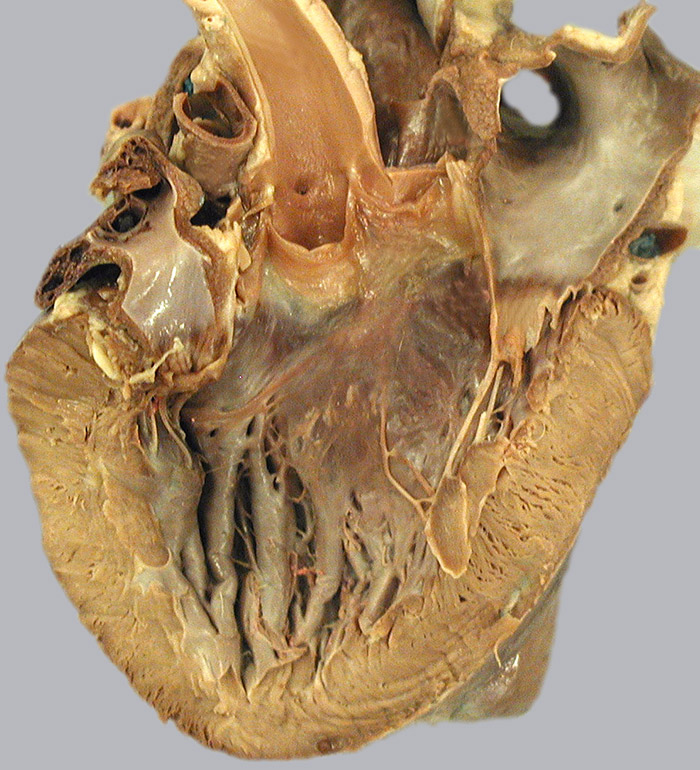
Isolated heart. A vertical incision has bisected the left atrium, the left atrioventricular opening and the left ventricle. Notice the wall thicknesses of the bisected left atrium (1) and left ventricle (2). Cusps (3) of the bisected left atrioventricular opening are connected by chordae tendineae (4) to a papillary muscle (5) and the ventricular wall. Ridges (6) lining the ventral ventricular wall are called trabeculae carnae. The ascending aorta has been opened, revealing the three semilunar cusps (7) of the aortic valve and the opening of the right coronary artery (8). The pulmonary trunk (9) and right & left side pulmonary veins (10) are evident. Notice the circumflex artery (11) and great cardiac vein (12) in the coronary groove.