
Lab 11 - Image 9
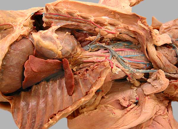
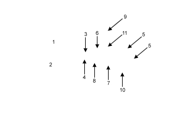
Cat dissection, left ventrolateral view of the thorax and neck. The heart (1) and left lung (2) are exposed. The aortic arch (not clearly visible) gives off the brachiocephalic trunk (3) and then the left subclavian a. (4). The brachiocephalic trunk gives off left and right common carotid arteries (5) and ends as a right subclavian a. (6). Before terminating as an axillary a. (7), each subclavian a. gives off four branches: costocervical trunk (8), internal thoracic a. (9), superficial cervical a. (10), and vertebral a. (not visible).
Notice the union of right and left brachiocephalic veins forming the cranial vena cava (11).