
Lab 11 - Image 2
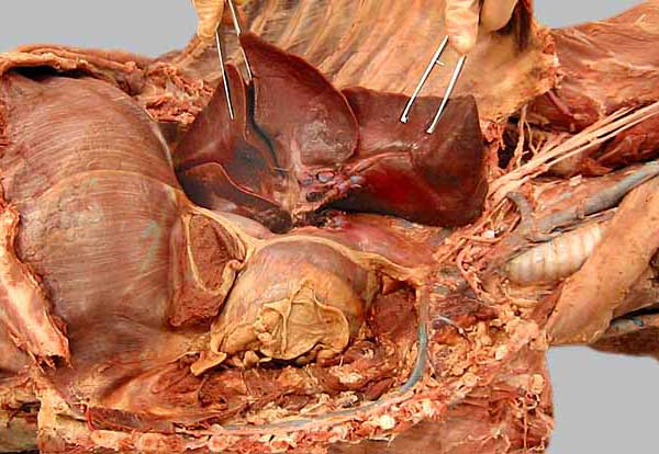
Caudal (1), middle (2), and cranial (3) lobes of the right lung are elevated and the root (4) of the lung has been cut to reveal the accessory lobe (5) of the right lung. The ventral part of the accessory lobe is hidden in a pocked formed by plica vena cava (6), which extends between the mediastinum (7) and the caudal vena cava (8). Also notice the heart (9), cranial vena cava (10), thymus (11), and phrenic n. (12).