
Lab 11 - Image 11
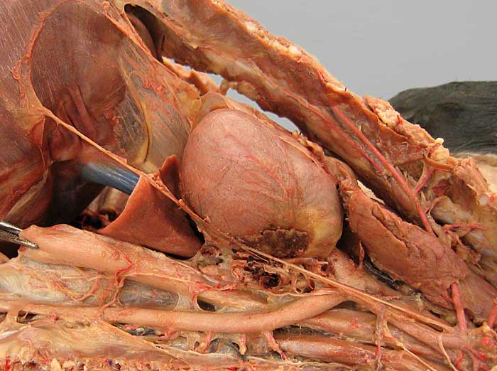
Cat dissection, left dorsolateral view of the thorax. The heart (1), accessory lobe of the right lung (2), and the thymus (3) are evident, as are the ascending aorta (4), the aortic arch (5) and the descending aorta (6). The latter gives rise to dorsal intercostal arteries (arrows). In this specimen, two bronchoesophageal arteries (7) can be seen supplying the esophagus (8) and the root of the left lung (9).
Also notice the caudal vena cava (10), the phrenic n. (11) innervating the diaphragm, and the internal thoracic a. (12).