
Lab 11 - Image 10
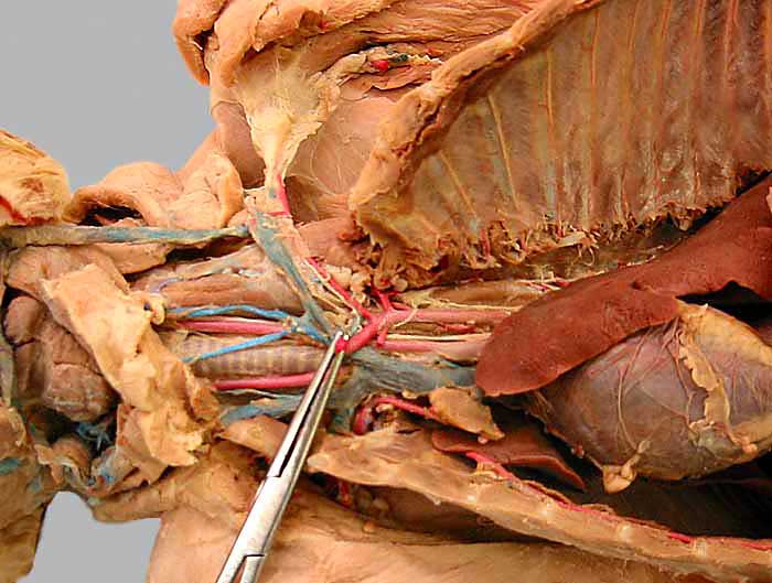
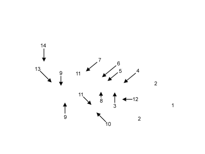
Cat dissection, ventral view of the cranial thorax and neck. The heart (1) and lungs (2) are evident. The aortic arch (not visible) gives off the brachiocephalic trunk (3) followed by the left subclavian a. (4). The latter gives off four branches: vertebral a. (5), costocervical trunk (6), superficial cervical a. (7), and internal thoracic a. (8), before terminating in the axillary a. (pulled medially by forceps). The brachiocephalic trunk gives off left and right common carotid arteries (9) and ends as a right subclavian a. (10).
Notice the union of right and left brachiocephalic veins (11) forming the cranial vena cava (12). Internal (13) and external jugular veins (14) are visible.