
Lab 10 - Image 9
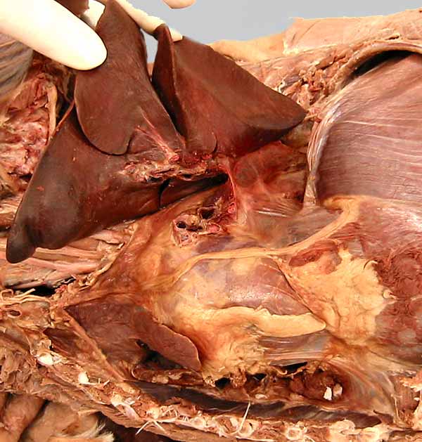

Thoracic viscera: The reflected left lung is composed of a cranial lobe with cranial (1) and caudal (2) parts and a caudal (3) lobe. The lung is covered by visceral (pulmonary) pleura. Connecting pleura, the pulmonary ligament (4), connects between visceral and parietal pleura. The mediastinum, including the heart (5), is covered by mediastinal parietal pleura. The diaphragm (6) is covered by diaphagmatic parietal pleura. The root of the lung (7) has been cut. The hilus of the lung (8), where the root joins the lung, is visible. The phrenic nerve (9) innervates the diaphragm.