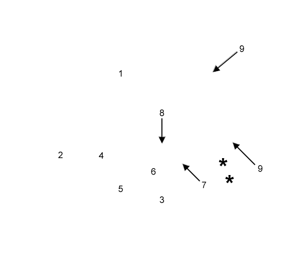
Lab 10 - Image 7


Cat dissection, right pleural cavity. The elevated thoracic wall (1) is covered by (transparent) costal parietal pleura. The diaphragm (2) is covered by diaphagmatic parietal pleura. The mediastinum, including the heart (3), is covered by mediastinal parietal pleura. The caudal (4), middle (5), and cranial (6) lobes of the right lung are covered by visceral (pulmonary) pleura. The right lung is reflected to reveal: phrenic n. (7), thymus (asterisk), azygos vein (8), and the internal thoracic a. (9).
