
Lab 10 - Image 10
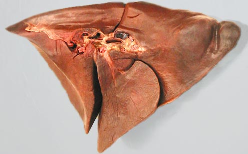
Isolated left lung, medial view. The left lung consists of a cranial lobe with cranial (1) and caudal (2) parts and a caudal (3) lobe. The pulmonary ligament (4) can be seen extending caudal to the hilus (5) of the lung. Lobar bronchi (6) go to each lobe of the lung. Pulmonary veins (7) contain red latex. Pulmonary arteries (8) contain blue latex.