
Lab 9 - Image 14
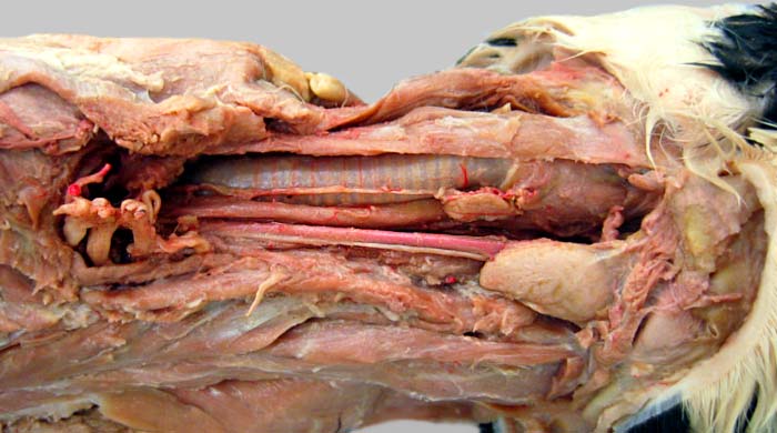
Neck, left lateral view, deep dissection; the head is to the right. After removing carotid sheath fascia, the vagosympathetic nerve trunk (1) can be seen attached to the common carotid a. (2). A recurrent laryngeal n. (3) is evident on the trachea (4). Other visible structures include: brachial plexus nerves (asterisks), esophagus (5), thyroid gland (6) and medial retropharyngeal lymph node (7).